Diagram Of Shoulder And Arm : Muscles Of The Pectoral Girdle And Upper Limbs Anatomy And Physiology - The shoulder plays a key role in the blood flow to the arms.
Diagram Of Shoulder And Arm : Muscles Of The Pectoral Girdle And Upper Limbs Anatomy And Physiology - The shoulder plays a key role in the blood flow to the arms.. It joins with the scapula above at the shoulder joint (or glenohumeral joint) and with the ulna and radius below at the elbow joint. If inflammation or an injury in the rotator cuff is present, this impingement causes pain. There are actually four joints that make up the shoulder. Sechrest, md narrates an animated tutorial on the basic anatomy of the shoulder. The upper arm includes the shoulder as well as the area between the shoulder and elbow joint.
The bones of the upper arm include the: The shoulder is the region where the upper limb is attached to the trunk. Subscapularis, supraspinatus, infraspinatus and teres minor. The muscles of the upper arm are responsible for the flexion and extension of the forearm at the elbow joint. The roof of the shoulder is formedby a part of the scapula called the acromion.
Shoulder Arm Hand Muscles Diagram Quizlet from o.quizlet.com There are actually four joints that make up the shoulder. Related posts of arm muscles diagram. The socket of the shoulder joint is shallow, and the labrum gives the socket more depth, and thus more stability. Related posts of diagram of shoulder muscles and tendons neck muscle anatomy mri. Is the wear and tear of shoulder cartilage until bare bone is exposed. To further reinforce the shoulder, the four muscles of the rotator cuff extend from the scapula and surround the head of the humerus to both rotate the arm and prevent dislocation. The roof of the shoulder is formedby a part of the scapula called the acromion. Also superior lateral cutaneous nerve of arm.;
What are common rotator cuff injuries?
It joins with the scapula above at the shoulder joint (or glenohumeral joint) and with the ulna and radius below at the elbow joint. Four of them are found on the anterior aspect of the shoulder, whereas the rest are located on the shoulder's posterior aspect and in the back. Neck muscle anatomy mri 12 photos of the neck muscle anatomy mri neck muscle anatomy images, neck muscle anatomy pictures, neck muscle anatomy posterior, neck muscle anatomy ultrasound, neck muscles anatomy radiology, human muscles, neck muscle anatomy images, neck muscle anatomy pictures, neck muscle anatomy. Diagram of the shoulder, including the location of the rotator cuff. Four muscles—the supraspinatus, infraspinatus, teres minor, and subscapularis. The main shoulder joint, called the glenohumeral joint, is formed The main shoulder muscles are trapezius, deltoid, pectoralis major and 4 rotator cuff muscles: The upper arm includes the shoulder as well as the area between the shoulder and elbow joint. Soft shoulder and varied terrain. Arthritis pain can occur at any time of day and can be present with or without shoulder stiffness. It rotates the forearm and also flexes the elbow. It can be present in the front, side or back of the shoulder. Sechrest, md narrates an animated tutorial on the basic anatomy of the shoulder.
When the arm is spun so that the thumb point to the outside of the body, meaning the palm of the hand looks forward then it is said the hand is supinated.but when the thumb remains in the inside and the palm. The armpit and shoulder serve as the meeting place for the torso and arms, so major vessels close to the heart travel through these areas. The shoulder plays a key role in the blood flow to the arms. Also superior lateral cutaneous nerve of arm.; Diagram of the shoulder, including the location of the rotator cuff.
Bones Of The Arm And Shoulder 417185 Vector Art At Vecteezy from static.vecteezy.com The armpit and shoulder serve as the meeting place for the torso and arms, so major vessels close to the heart travel through these areas. A dislocated shoulder occurs when the humerus (upper arm bone) separates from the shoulder blade at the main shoulder joint. The bones of the shoulder are: Neck muscle anatomy mri 12 photos of the neck muscle anatomy mri neck muscle anatomy images, neck muscle anatomy pictures, neck muscle anatomy posterior, neck muscle anatomy ultrasound, neck muscles anatomy radiology, human muscles, neck muscle anatomy images, neck muscle anatomy pictures, neck muscle anatomy. The acromioclavicular joint is formed by an articulation between the lateral end of the clavicle and the acromion process of the scapula. From the arm muscle diagram above, the muscles of the arm that can be seen easily on the surface include biceps, triceps, brachioradialis, extensor carpi radialis longus, and deltoid.biceps are large muscle of the upper arm is formally known as the biceps brachii muscle, and rests on top of the humerus bone. If the frozen shoulder syndrome is present, the painful arm will not rotate outward in comparison to the healthy shoulder. Shoulder muscles move the shoulder blades and upper arm bones.
See more ideas about muscle anatomy, anatomy, shoulder muscle anatomy.
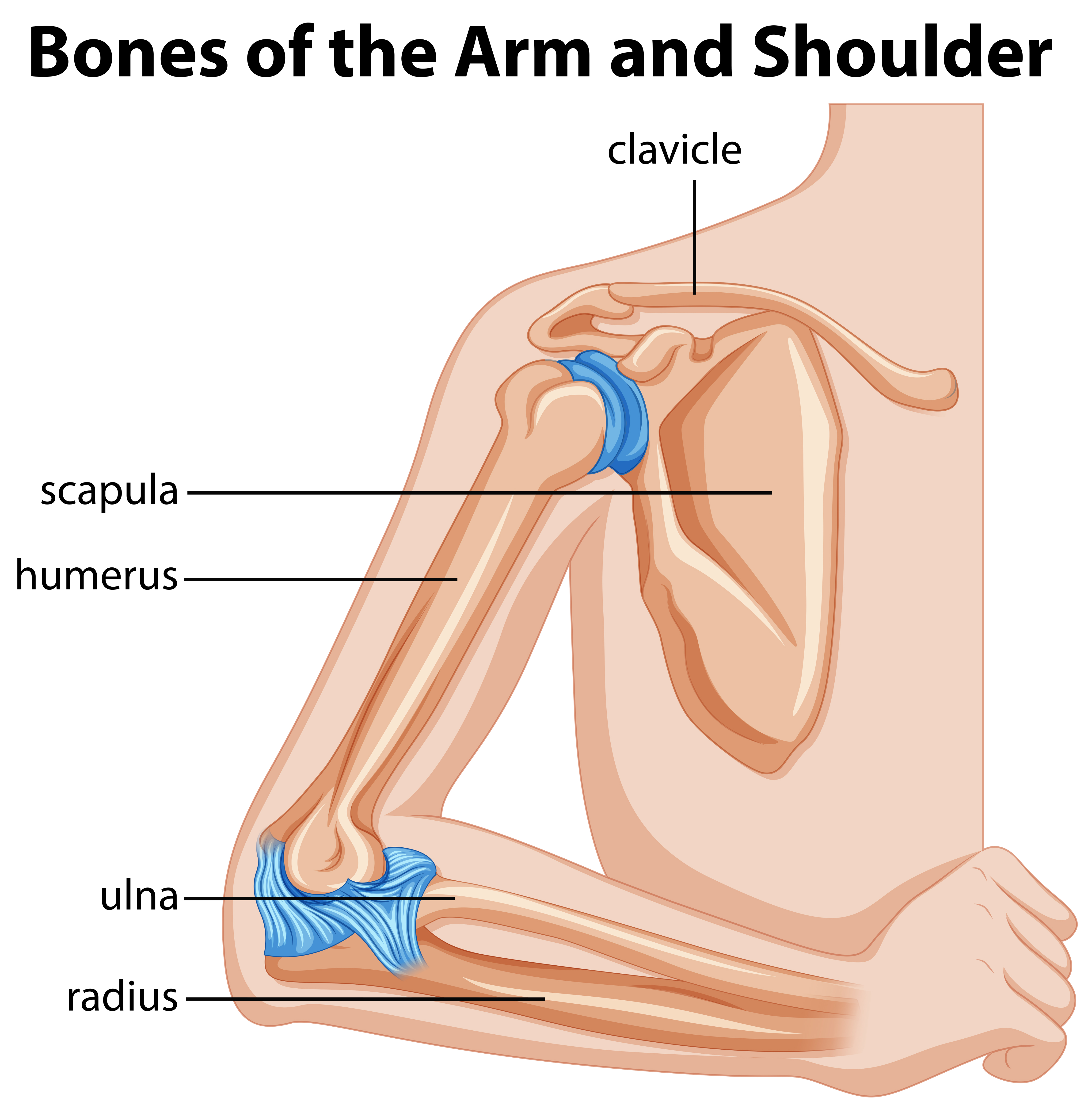
Some people have pain even when they are not using the arm, and some have pain only when using it. Test your knowledge of the clavicle, scapula and humerus with our labeled diagram exercises and quizzes! The shoulder isn't just one bone, it's actually made up of three different bones and various tendons, ligaments, and muscles.the three bones located in the shoulder are the humerus, the scapula, and the clavicle. Shoulder muscles move the shoulder blades and upper arm bones. The simplest approach to read a home wiring diagram is to begin at the source, or the major power supply. Four of them are found on the anterior aspect of the shoulder, whereas the rest are located on the shoulder's posterior aspect and in the back. You can see in the arm muscle diagram above that there are important parts in arm muscles. The bones of the shoulder are: Subscapularis, supraspinatus, infraspinatus and teres minor. Each time the arm is raised, not only does the ball of the humerus move in the socket of the. There are actually four joints that make up the shoulder. From the arm muscle diagram above, the muscles of the arm that can be seen easily on the surface include biceps, triceps, brachioradialis, extensor carpi radialis longus, and the arm muscles of the upper limb act on the elbow and shoulder joints to produce the various movements of the forearm. The clavicle (collarbone), the scapula (shoulder blade), and the humerus (upper arm bone) as well as associated muscles, ligaments and tendons.
Externally rotate the arms outward. Shoulder joint injuries can be head. Soft shoulder and varied terrain. Some people have pain even when they are not using the arm, and some have pain only when using it. What are common rotator cuff injuries?
Labeled Human Anatomy Diagram Of Man S Arm Shoulder And Upper Back Stock Images Page Everypixel from media.istockphoto.com The humerus is the bone of the arm that articulates with. The arm is one of the body's most complex and frequently used structures. What are common rotator cuff injuries? The largest bone of the arm, the humerus connects to the scapula and clavicle in the shoulder. Shoulder and arm (labeled) 1 of 1 human science human body arm anchor chart bone shoulder health skeleton diagram Neck muscle anatomy mri 12 photos of the neck muscle anatomy mri neck muscle anatomy images, neck muscle anatomy pictures, neck muscle anatomy posterior, neck muscle anatomy ultrasound, neck muscles anatomy radiology, human muscles, neck muscle anatomy images, neck muscle anatomy pictures, neck muscle anatomy. When the arm is spun so that the thumb point to the outside of the body, meaning the palm of the hand looks forward then it is said the hand is supinated.but when the thumb remains in the inside and the palm. A dislocated shoulder occurs when the humerus (upper arm bone) separates from the shoulder blade at the main shoulder joint.
The bones of the shoulder are:
Shoulder and arm (labeled) 1 of 1 human science human body arm anchor chart bone shoulder health skeleton diagram There are actually four joints that make up the shoulder. Shoulder muscles move the shoulder blades and upper arm bones. Pain in the shoulder joint is the major sign of arthritis. Is the wear and tear of shoulder cartilage until bare bone is exposed. The humerus is the bone of the arm that articulates with the scapula proximally and with the radius and the ulna distally. In this episode of eorthopodtv, orthopaedic surgeon randale c. The list of muscles and their functions are presented below. Most relevant best selling latest uploads. The acromioclavicular joint is formed by an articulation between the lateral end of the clavicle and the acromion process of the scapula. If the frozen shoulder syndrome is present, the painful arm will not rotate outward in comparison to the healthy shoulder. A final test for frozen shoulder is to stand with both arms at the sides and the elbows flexed at ninety degrees. The shoulder plays a key role in the blood flow to the arms.
The armpit and shoulder serve as the meeting place for the torso and arms, so major vessels close to the heart travel through these areas diagram of shoulder. A final test for frozen shoulder is to stand with both arms at the sides and the elbows flexed at ninety degrees.


0 Komentar