Rib Cage Anatomy Posterior / Human Skeleton System Rib Cage Anatomy Posterior View Stock Photo Picture And Royalty Free Image Image 92995436 : The articulation with the rib cage leads to regional variations in movement patterns and function (1).
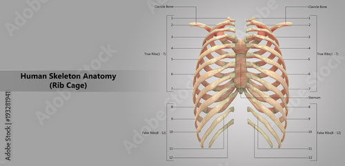
Rib Cage Anatomy Posterior / Human Skeleton System Rib Cage Anatomy Posterior View Stock Photo Picture And Royalty Free Image Image 92995436 : The articulation with the rib cage leads to regional variations in movement patterns and function (1).. The ribs are a set of twelve paired bones which form the protective 'cage' of the thorax. It branches from the ileocolic artery and may branch further to the appendicular artery. They articulate with the vertebral column posteriorly, and terminate anteriorly as cartilage (known as costal cartilage). A rib has a flat body, as you can see from the picture of the anatomy of the human rib cage. There are twelve (12) pairs of ribs and all articulate posteriorly with the thoracic vertebrae.
Anatomy the rib cage is a bony structure found in the chest (thoracic cavity). Of these, 35% were cervical ribs. The angles of the ribs form the most posterior extent of the thoracic cage. These pass from the inferior edge of the costal groove to the superior margins of the ribs below. Gross anatomy there are 12 pairs of ribs which are separated by intercostal spaces.
Ribcage Musculoskeletal Key from i1.wp.com The ribs are attached to corresponding thoracic vertebrae posteriorly. They articulate with the vertebral column posteriorly, and terminate anteriorly as cartilage (known as costal cartilage). Anatomy the rib cage is a bony structure found in the chest (thoracic cavity). Rib cage, in vertebrate anatomy, basketlike skeletal structure that forms the chest, or thorax, and is made up of the ribs and their corresponding attachments to the sternum (breastbone) and the vertebral column. Related posts of rib cage diagram with organs woman stomach anatomy. Anteriorly, most are attached directly to the sternum. A rib has a flat body, as you can see from the picture of the anatomy of the human rib cage. The flexible (hyaline) cartilage, makes the breathing process easier.
There are twelve (12) pairs of ribs and all articulate posteriorly with the thoracic vertebrae.
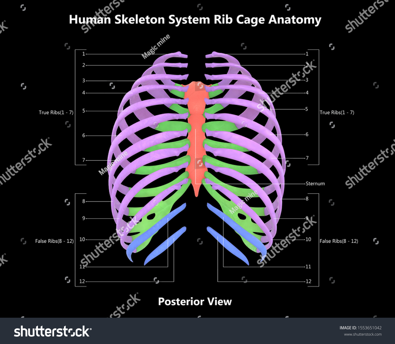
Of these, 35% were cervical ribs. Lateral view of a pair of ribs articulating with the thoracic vertebrae. The angles of the ribs form the most posterior extent of the thoracic cage. There are twelve (12) pairs of ribs and all articulate posteriorly with the thoracic vertebrae. Rib cage anatomy posterior human skeleton system skelett anatomie menschliches bone scheletro sistema knochen thoracic menselijk humain anatomia maenskligt umano. Extent of the region and the articulations with the rib cage. Posteriorly, the heads of the ribs interdigitate with the vertebrae and are numbered according. Muscle anatomy drawing 12 photos of the muscle anatomy drawing anatomy muscle sketches, arm. The ribs are attached to corresponding thoracic vertebrae posteriorly. The articulation with the rib cage leads to regional variations in movement patterns and function (1). The primary responsibilities of the ribcage involve protecting the thoracic visceral organs, enclosing the thoracic visceral organs, and is included. The flexible (hyaline) cartilage, makes the breathing process easier. Rib cage pain can be caused.
The angles of the ribs form the most posterior extent of the thoracic cage. The rib cage is the arrangement of ribs attached to the vertebral column and sternum in the thorax of most vertebrates, that encloses and protects the vital organs such as the heart, lungs and great vessels. It branches from the ileocolic artery and may branch further to the appendicular artery. Anatomy the rib cage is a bony structure found in the chest (thoracic cavity). The eleven pairs of internal intercostal muscles are found posterior to the external intercostals.
Human Skeleton System Rib Cage With Detailed Labels Anatomy Posterior View Stock Illustration Adobe Stock from as2.ftcdn.net At the chest, many rib bones connect to the sternum via costal cartilage,. The rib cage is collectively made up of long, curved. The ribs are attached to corresponding thoracic vertebrae posteriorly. Related posts of rib cage diagram with organs woman stomach anatomy. The primary responsibilities of the ribcage involve protecting the thoracic visceral organs, enclosing the thoracic visceral organs, and is included. A rib has a flat body, as you can see from the picture of the anatomy of the human rib cage. In this video, we explore:1) the anatomy of the sternum2) the anatomy and differences between the three classes of ribs3) the anatomy and differences between. It branches from the ileocolic artery and may branch further to the appendicular artery.
The rib cage is collectively made up of long, curved.
The flexible (hyaline) cartilage, makes the breathing process easier. It may occur after an obvious injury or without explanation. In this video, we explore:1) the anatomy of the sternum2) the anatomy and differences between the three classes of ribs3) the anatomy and differences between. The nomenclature of the costal veins is the same as the arteries. The primary responsibilities of the ribcage involve protecting the thoracic visceral organs, enclosing the thoracic visceral organs, and is included. At the chest, many rib bones connect to the sternum via costal cartilage,. In the inferior pair of ribs (i), the posterior rib (arrow) is slightly lower than the anterior rib. Its function is to elevate the ribs. The first rib is attached to thoracic vertebra. The posterior abdominal wall is a musculoskeletal structure formed by the posterior abdominal muscles posteriorly by the lumbar vertebrae, muscles, and fascia. The rib cage is collectively made up of long, curved. On the interior wall of the rib body is a channel, sulcus costae, with blood vessels and nerves. As part of the bony thorax, the ribs protect the internal thoracic organs.
Rib cage, in vertebrate anatomy, basketlike skeletal structure that forms the chest, or thorax, and is made up of the ribs and their corresponding attachments to the sternum (breastbone) and the vertebral column. The nomenclature of the costal veins is the same as the arteries. The first rib is attached to thoracic vertebra. Its function is to elevate the ribs. Costae) are long, flat, curved bones that form the rib cage.there are twelve pairs of ribs, all of which articulate with the vertebral column, while only the first seven ribs directly articulate with the sternum.the rib cage forms the majority of the thoracic skeleton and provides protection for the internal thoracic organs, including the lungs and the heart.
Human Skeleton System Rib Cage Detailed Stock Illustration 1553651042 from image.shutterstock.com The eleven pairs of internal intercostal muscles are found posterior to the external intercostals. The rib cage is collectively made up of long, curved. However, they do not attach directly to the sternum anteriorly, and instead, attach to the. Rib cage pain can be caused. In the anatomical position, the angles align with the medial border of the scapula. The rib cage, also distinguished as the thoracic cage, is a bony and cartilaginous structure which forming a core portion of the human skeleton. Rib cage anatomy the rib cage, shaped in a mild cone shape and more flexible than most bone sets, is made up of varying elements such as the thoracic vertebra, 12 equally paired ribs, costal cartilage, and held together anteriorly by the sternum. Ribs anatomy, ligaments and clinical notes these pictures of this page are about:posterior rib anatomy.
In the inferior pair of ribs (i), the posterior rib (arrow) is slightly lower than the anterior rib.
Lateral view of a pair of ribs articulating with the thoracic vertebrae. At the chest, many rib bones connect to the sternum via costal cartilage,. Lateral view of a pair of ribs articulating with the thoracic vertebrae. Anteriorly, most are attached directly to the sternum. Measuring rib cage and abdominal movement is the most common technique for assessing respiratory effort in laboratory. The upper edge is round and the lower sharp. In the inferior pair of ribs (i), the posterior rib (arrow) is slightly lower than the anterior rib. Its function is to elevate the ribs. The primary responsibilities of the ribcage involve protecting the thoracic visceral organs, enclosing the thoracic visceral organs, and is included. The angles of the ribs form the most posterior limit of the thoracic cage and are in line with the medial border of the scapula. Rib cage, in vertebrate anatomy, basketlike skeletal structure that forms the chest, or thorax, and is made up of the ribs and their corresponding attachments to the sternum (breastbone) and the vertebral column. Rib cage anatomy posterior human skeleton system skelett anatomie menschliches bone scheletro sistema knochen thoracic menselijk humain anatomia maenskligt umano. Posterior view of vertebrae anatomy.
The angles of the ribs form the most posterior extent of the thoracic cage rib cage anatomy. The shape of the thoracic cage is like a domed bird cage with the horizontal bars formed by ribs and their associated costal cartilages.


0 Komentar